

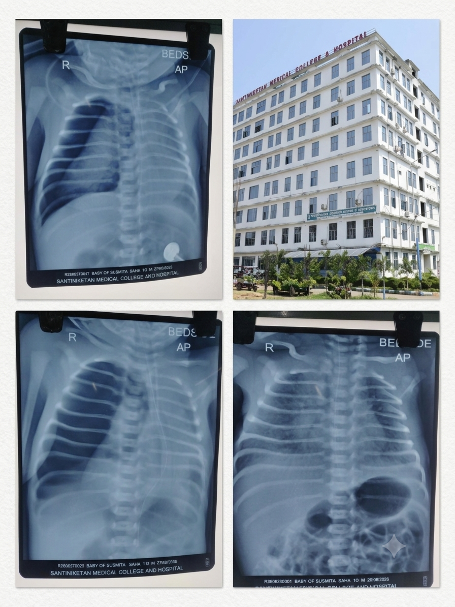
Premature Newborn Receives Emergency Life-Saving Treatment at Santiniketan Medical College & Hospital
34-Week-Old Baby Develops Severe Respiratory Complications; Timely Diagnosis and NICU-Based Emergency Intervention Bring Encouraging Improvement
A premature male newborn received critical emergency treatment at Santiniketan Medical College & Hospital after developing severe respiratory complications within minutes of birth. The baby, born to Mrs. Sushmita Das, a resident of Bolpur in Birbhum district, required intensive neonatal care following his premature delivery at 34 weeks of gestation.
What initially appeared as respiratory distress soon after birth gradually became more complicated. The newborn required oxygen support and subsequently Continuous Positive Airway Pressure (CPAP) when his breathing difficulty increased and his blood oxygen saturation began to decline.
During the course of treatment, a chest X-ray indicated findings suggestive of pneumonia. The baby was therefore managed with appropriate antibiotics, intravenous fluids and continued respiratory support under close observation in the Neonatal Intensive Care Unit (NICU).
The situation became particularly critical on the morning of 27 August 2026, when the baby’s oxygen saturation suddenly began to fall. A repeat chest X-ray revealed a right-sided pneumothorax. The medical team immediately initiated emergency treatment, first performing needle aspiration and subsequently deciding to insert an intercostal drainage (ICD) tube when the pneumothorax showed only partial resolution.
The ICD procedure was performed at the NICU bedside by hospital surgeon Dr. Javid V. Siddiqui at approximately 5:00 PM on 27 August, in the presence of doctors from the Paediatrics Department and under the advice of Prof. (Dr.) Asok Kumar Datta.
Following the emergency intervention, the baby’s respiratory distress reduced significantly. As of approximately 9:00 PM on 27 August, the newborn was showing encouraging improvement and was maintaining blood oxygen saturation close to 100 percent with ongoing respiratory support.
Step 1: Premature Birth Created an Immediate Medical Challenge
The baby was delivered by Caesarean section on 25 August 2026 at approximately 9:00 PM.
Born at only 34 weeks of gestation, the newborn required special medical attention immediately after delivery. Premature babies may require intensive monitoring because their developing organs, including the respiratory system, may not yet be fully mature.
In this case, the newborn developed respiratory distress within only a few minutes of birth. Recognising the need for specialised neonatal care, the medical team promptly shifted the baby to the hospital’s NICU.
The decision to move the newborn immediately into an intensive care environment allowed doctors and nursing staff to continuously monitor his condition and respond quickly to changes in his breathing and oxygen levels.
Step 2: Oxygen Support Was Started Immediately
After admission to the NICU, the newborn was initially provided with oxygen support.
The medical team closely observed his respiratory pattern and blood oxygen saturation. Despite the initial respiratory assistance, however, the baby’s breathing difficulty gradually increased.
At the same time, his oxygen saturation began to decline. This indicated that the existing respiratory support was no longer sufficient to adequately manage his condition.
Doctors therefore decided to escalate the respiratory support while continuing close monitoring.
Step 3: CPAP Support Was Introduced
As the newborn’s respiratory distress worsened, the medical team placed him on Continuous Positive Airway Pressure, or CPAP.
CPAP is a form of non-invasive respiratory support that provides a continuous flow of air under controlled pressure. In neonatal care, it can be used to assist babies who are experiencing difficulty maintaining effective breathing.
For this premature newborn, CPAP became an important component of his treatment as the medical team attempted to stabilise his breathing and maintain adequate oxygenation.
The baby remained under continuous observation in the NICU while the doctors assessed his response to the treatment.
Step 4: Chest X-Ray Suggested Pneumonia
As respiratory difficulties continued, the medical team conducted a chest X-ray to assess the condition of the newborn’s lungs.
The X-ray findings were suggestive of pneumonia.
Following the investigation, the newborn’s treatment was continued with appropriate antibiotics. Intravenous fluids were also administered, while respiratory support remained ongoing.
At this stage, the baby’s condition was described as relatively stable. However, because of his premature birth and continuing respiratory problems, the medical team maintained close observation.
The NICU team continued to watch for any sudden changes that might indicate a new complication.
Step 5: A Sudden Deterioration on 27 August
The situation changed on the morning of 27 August 2026.
The newborn’s blood oxygen saturation began to fall suddenly. Such an unexpected decline in a critically ill premature infant required immediate clinical assessment.
The treating doctors examined the baby and suspected that a new complication could be responsible for the sudden deterioration.
Without delay, they ordered a repeat chest X-ray.
The investigation proved crucial in identifying the cause of the baby’s worsening respiratory condition.
Step 6: Right-Sided Pneumothorax Was Detected
The repeat chest X-ray revealed a right-sided pneumothorax.
Pneumothorax occurs when air collects in the pleural space surrounding the lung. The accumulation of air can interfere with normal lung expansion and may lead to worsening respiratory distress and reduced oxygenation.
For a premature newborn who was already dependent on respiratory support, the development of pneumothorax represented a serious medical emergency.
The medical team immediately moved to address the problem.
The priority was to remove the accumulated air and improve the baby’s ability to breathe and maintain adequate oxygen levels.
Step 7: Emergency Needle Aspiration Was Performed
The doctors first performed needle aspiration to remove the accumulated air from the pleural cavity.
A significant amount of air was successfully aspirated.
Following the procedure, the baby’s condition showed some initial improvement. However, the improvement was not complete.
His oxygen saturation continued to fluctuate, indicating that the underlying problem had not yet been fully resolved.
The medical team therefore continued close observation and conducted another chest X-ray to assess the response to the emergency intervention.
Step 8: Further Imaging Showed Only Partial Resolution
The repeat chest X-ray showed that the pneumothorax had improved only partially.
This finding was significant because it suggested that air was continuing to accumulate or that the existing collection had not been completely relieved.
Given the baby’s critical condition and the concern regarding a persistent air leak, the treating doctors concluded that a more definitive drainage method was required.
The medical team decided that an intercostal drainage (ICD) tube should be inserted.
The purpose of the procedure was to allow continuous drainage of air from the pleural space and help stabilise the newborn’s respiratory condition.
Step 9: Surgeon Was Called for the Emergency Procedure
Because the newborn required an urgent surgical intervention, hospital surgeon Dr. Javid V. Siddiqui was immediately called.
At that point, the baby was critically ill and remained dependent on intensive neonatal support.
The treating team considered the risks associated with shifting such a fragile newborn away from the NICU. Since respiratory support and continuous monitoring were already being provided inside the NICU, it was decided that the emergency procedure should be performed at the bedside.
This approach allowed the newborn to remain in the controlled environment of the NICU throughout the intervention.
Step 10: ICD Procedure Was Successfully Performed at the NICU Bedside
At approximately 5:00 PM on 27 August 2026, Dr. Javid V. Siddiqui performed the intercostal drainage procedure.
The procedure was carried out in the presence of doctors from the Paediatrics Department and under the advice of Prof. (Dr.) Asok Kumar Datta.
The decision to perform the procedure directly inside the NICU was based on the newborn’s critical condition and the need to maintain immediate access to respiratory support and continuous monitoring.
The ICD tube provided a means for continued drainage of air from the pleural space.
The medical team continued monitoring the newborn closely following the procedure.
Step 11: Respiratory Distress Reduced After the Intervention
Following the emergency ICD procedure, the baby’s respiratory distress reduced significantly.
The improvement represented an encouraging development after a prolonged period of respiratory instability.
The drainage tube remained in place, while the newborn continued to receive intensive care in the NICU.
Doctors and nursing staff maintained continuous observation to monitor his respiratory status and oxygen saturation and to assess his response to ongoing treatment.
The baby continued to receive respiratory support as required.
Step 12: Oxygen Saturation Improved Significantly
By approximately 9:00 PM on 27 August 2026, the newborn had shown encouraging improvement.
With continued respiratory support, his blood oxygen saturation was being maintained close to 100 percent.
For the treating team, this represented a positive response following the emergency intervention.
However, despite the improvement, the newborn continued to require intensive neonatal care because of his premature birth and the severity of the complications he had experienced.
The medical team therefore continued to exercise caution and maintained close monitoring.
Step 13: Intensive Treatment Continues
The emergency intervention was an important step in stabilising the baby’s condition, but his treatment did not end with the insertion of the drainage tube.
The newborn continues to receive intensive care in the NICU.
His treatment includes appropriate antibiotics, intravenous fluids, respiratory support and continuous monitoring.
The medical team is closely observing his clinical condition and response to treatment. Particular attention is being given to his respiratory status and oxygenation.
The drainage tube remains in place while doctors continue to assess the baby’s progress.
Further management will depend on his clinical response and subsequent investigations.
Step 14: Parents Kept Informed Throughout the Treatment
Throughout the course of treatment, the newborn’s parents were informed about his condition, investigations and treatment.
The medical team counselled the family at different stages as the baby’s condition changed.
For the parents, the sudden respiratory deterioration and subsequent diagnosis of pneumothorax represented a highly stressful situation. Clear communication regarding the baby’s medical condition and the need for emergency intervention was therefore an important part of the overall care process.
The family was kept aware of the baby’s progress following the emergency procedure.
Step 15: Coordinated Medical Care Played a Key Role
The newborn’s treatment involved several successive stages, with the medical team responding to changes in his condition as they occurred.
The sequence began with immediate NICU admission following respiratory distress after premature birth.
Oxygen support was followed by CPAP when respiratory distress increased. A chest X-ray subsequently suggested pneumonia, leading to antibiotic therapy, intravenous fluids and continued respiratory support.
When oxygen saturation suddenly declined on 27 August, the medical team promptly investigated the change. The repeat X-ray revealed right-sided pneumothorax.
Emergency needle aspiration was performed, but when the pneumothorax showed only partial resolution and oxygen levels continued to fluctuate, the team escalated treatment to intercostal drainage.
The successful bedside ICD procedure required coordination between the surgical and Paediatrics teams, along with continued NICU support.
Step 16: NICU Care Remains Essential
The case highlights the importance of specialised neonatal intensive care for premature babies who develop serious respiratory complications.
A premature newborn may require continuous monitoring and respiratory assistance, particularly when breathing difficulties develop soon after delivery.
In this case, the availability of NICU-based respiratory support allowed the medical team to respond rapidly to changes in the baby’s condition.
The sudden fall in oxygen saturation was investigated immediately, and the underlying pneumothorax was identified through repeat imaging.
The ability to perform the emergency drainage procedure within the NICU also allowed the baby to remain under continuous respiratory and clinical monitoring.
Step 17: Improvement Brings Hope, but Caution Remains Necessary
The baby’s response following emergency intervention has provided a positive sign for his ongoing treatment.
The reduction in respiratory distress and improvement in oxygen saturation following ICD drainage are encouraging developments.
However, the newborn remains critically ill and continues to require intensive neonatal care.
His premature birth, recent pneumothorax and ongoing need for respiratory support mean that his condition must continue to be monitored carefully.
The medical team therefore remains cautiously optimistic rather than declaring the baby completely out of danger.
A Critical 48-Hour Journey Under Intensive Medical Supervision
From the moment of his birth on 25 August to the emergency intervention on 27 August, the newborn went through a complex medical journey.
The first challenge was premature birth at 34 weeks, followed within minutes by respiratory distress. The baby was promptly shifted to the NICU and provided with oxygen support.
As his breathing difficulty increased and oxygen saturation fell, CPAP support was initiated.
A subsequent chest X-ray suggested pneumonia, leading to treatment with appropriate antibiotics, intravenous fluids and continued respiratory assistance.
For a period, the newborn remained relatively stable. However, on the morning of 27 August, his oxygen saturation suddenly declined.
The medical team responded immediately. A repeat chest X-ray revealed a right-sided pneumothorax.
Emergency needle aspiration removed a significant amount of accumulated air and initially improved the baby’s condition. But continuing fluctuations in oxygen saturation and only partial resolution on follow-up imaging indicated that additional intervention was necessary.
The decision was then taken to place an ICD tube.
Dr. Javid V. Siddiqui was called for the emergency procedure. Considering the baby’s critical condition and the potential risks of moving him, the procedure was carried out directly at the NICU bedside.
At approximately 5:00 PM on 27 August, the intercostal drainage procedure was successfully performed in the presence of Paediatrics Department doctors and under the advice of Prof. (Dr.) Asok Kumar Datta.
Several hours later, at approximately 9:00 PM, the baby showed encouraging signs of improvement. His respiratory distress had reduced, and his oxygen saturation was being maintained close to 100 percent with ongoing respiratory support.
Medical Team Remains Cautiously Hopeful
The newborn’s journey is not yet complete. Although the emergency intervention has produced a significant improvement, he remains a critically ill premature infant requiring continued intensive care.
The drainage tube remains in place, and the baby continues to receive respiratory support, antibiotics, intravenous fluids and continuous monitoring.
The medical team at Santiniketan Medical College & Hospital is continuing to assess his condition carefully and provide ongoing neonatal care.
For the family, the improvement following the emergency intervention offers a measure of hope after a difficult and uncertain period.
At present, however, doctors remain cautious. The coming period of intensive monitoring will be important in determining the baby’s further recovery.
The case demonstrates how rapidly a premature newborn’s condition can change and how timely recognition of complications, immediate investigation and coordinated emergency management can become critical components of neonatal care.
For this 34-week-old baby, the successful emergency intervention has marked an encouraging turning point. With continued intensive care and close monitoring, the medical team remains cautiously hopeful about his recovery.
Read More: AI Ma: Antyodaya Digital Mission Brings Technology!!!





[…] Read More: Santiniketan Medical College & Hospital: Premature Newborn Receives Emergency Life-Saving Treatm… […]